Services
Common procedures and Services:
- Electrocardiogram (ECG or EKG)
- Echocardiography
- Transesophageal Echocardiogram (TEE)
- Transthoracic Echocardiogram (TTE)
- Exercise Stress Test (Regular Treadmill)
- Exercise Stress Test with Nuclear Imaging
- Pharmacological Stress Test
- Stress Echocardiogram
- Bubble Study
- Coronary Computed Tomography Angiogram (CTA)
- Implantable Cardioverter/Defibrillators and Pacemaker Interrogation
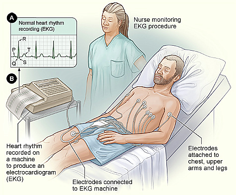
Electrocardiogram (ECG/EKG)
Electrocardiograms, abbreviated as ECGs or EKGs, play a crucial role in swiftly identifying heart issues and monitoring overall cardiac health. These tests capture the electrical signals within the heart, and they are commonly conducted in medical facilities like doctors’ offices, clinics, and hospital rooms. ECG machines are integral in operating rooms and ambulances.
Echocardiography
Echocardiography, commonly known as echo, is a noninvasive examination utilizing ultrasound to visualize the structures of the heart. This test generates dynamic images of the heart through the use of sound waves. These images provide insights into the size and shape of the heart, as well as its ability to pump blood effectively.
The echocardiogram is instrumental in identifying various conditions, including the presence of blood clots within the heart, accumulation of fluid in the pericardium (the sac surrounding the heart), detection of tumors, and assessment of issues with the aorta.
Transesophageal Echocardiogram (TEE)
A transesophageal echocardiogram (TEE) utilizing ultrasound enables your physician to obtain a more detailed view of your heart. In the course of this examination, a tube is introduced through your throat and carefully directed into your esophagus, the conduit connecting your mouth and stomach.

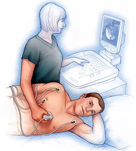
Transthoracic Echocardiogram (TTE)
A transthoracic echocardiogram, commonly known as TTE, stands as the prevailing form of echocardiography. This procedure involves affixing a transducer to your chest, following the application of a gel to your skin. The transducer releases ultrasound waves that traverse your chest wall, reaching your heart.
Within the echocardiogram examination, these ultrasound waves interact with the structures in your heart, translating them into visual images displayed on a screen.
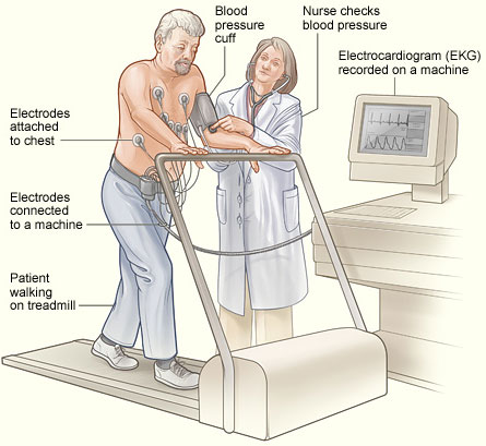
Exercise Stress Test (Regular Treadmill)
The exercise stress test, commonly referred to as a treadmill test, assesses the heart’s performance during physical activity, particularly while walking on a treadmill. As the body engages in more intense work, the heart pumps increased blood, demanding additional oxygen.
In stress echocardiography, ultrasound imaging is employed to evaluate the heart’s efficiency in pumping blood throughout the body. This diagnostic tool not only gauges cardiac function but also aids in tailoring exercise recommendations, specifying the type and intensity of physical activity suitable for an individual patient.
Exercise Stress Test with Nuclear Imaging
During a nuclear exercise stress test, a minute quantity of a radioactive tracer is introduced into a vein to assess blood circulation to the heart. Utilizing a gamma camera that detects the emitted radiation from the tracer, computer-generated images of the heart are generated. When combined with physical activity, this test aids in discerning whether the heart receives sufficient blood flow during exertion compared to periods of rest.
Bubble Study
Bubble echocardiograms employ a procedure akin to a regular echocardiogram, with the insertion of an intravenous (IV) line in the patient’s arm. At specific intervals in the imaging process, saline infused with bubbles is introduced into the vein.
This technique provides additional insights, enabling the identification of potential blood flow abnormalities within the heart. It proves particularly effective in pinpointing the existence of a hole in the heart, making it one of the most reliable methods for such diagnoses.

Coronary Computed Tomography Angiogram (CTA)
Cardiovascular computed tomography angiography (CTA), a noninvasive diagnostic procedure, employs X-rays to specifically target the coronary arteries. This imaging technique enables physicians to identify potential blockages in the arteries. To visualize the vessels in the heart, a dye (iodine-based contrast material) is administered through a vein in the arm.
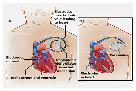
Implantable Cardioverter/Defibrillators and Pacemaker Interrogation
A cardioverter-defibrillator (ICD) is a chest-implanted device designed to electrically stimulate the heart in the presence of severe rhythm disturbances. Additionally, it may incorporate an internal pacemaker to address less severe irregularities in your heart rate.
Regular interrogations of both the pacemaker and ICD are conducted to oversee and manage abnormal heartbeats, ensuring proper blood flow throughout the body.